Technology Transfer
Licensing opportunities
3D co-culture of podocytes and endothelial cells
– Applications
- In vitro study model of pathologies affecting kidney glomerular permeability.
- Drug delivery tests (particularly to the renal glomerular filtration barrier).
- Drug toxicity assays.
– Key Benefits
- Effective 3D method for co-culturing podocytes and endothelial cells.
- Simple and inexpensive application.
– Offer
- Licensing out.
- Co-Development.
Invention
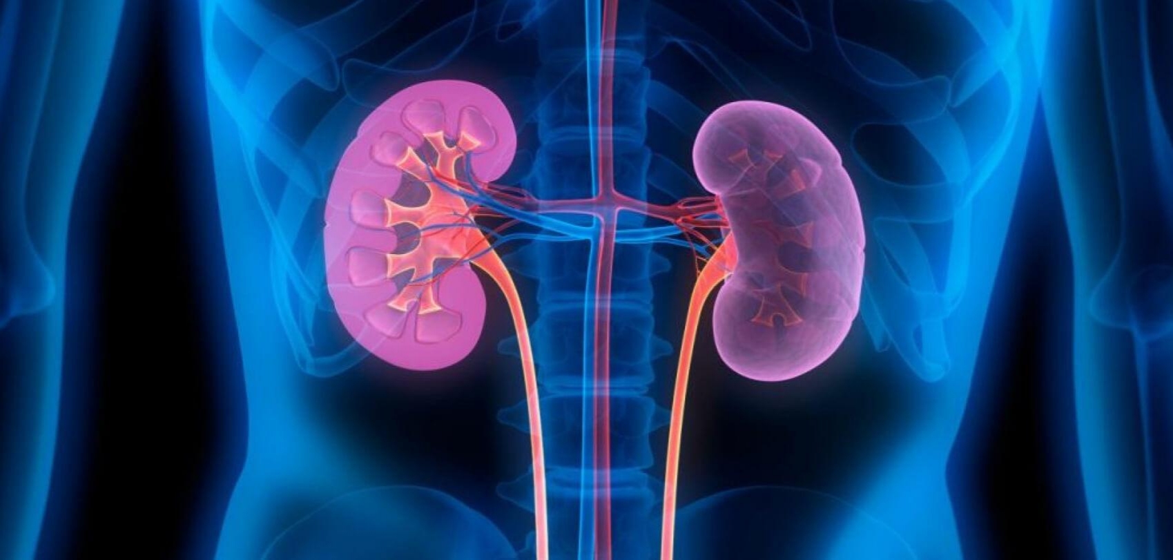
A new three-dimensional co-culture method of podocytes and endothelial cells for in vitro model study of pathologies affecting kidney and, in particular, the renal glomerular filtration barrier.
Background
Cells live in a 3D microenvironment and particularly, podocytes in the glomerulus make adhesions to the external side of the glomerular basement membrane, whereas endothelial cells line the internal side. Podocytes, endothelial cells and the intervening glomerular basement membrane form together the glomerular filtration barrier. The need for co-culturing podocytes and endothelial cells is widely recognized in the nephrology research community. This method is the first successful 3D system for coculturing them in vitro.
Technology
This method allows podocytes and endothelial cells to grow on a isoporous membrane, covering the external and internal side respectively. Thus, cells make adhesion only to the membrane itself, though on the opposite sides. Compared to artificial capillaries, this system is unexpensive and easier to use and does not require specific equipment. This method, as per in vivo situation, allows multiple analyses on both cell types. The presence of the membrane between the two cell types and the complete separation of their own supernatants allows to perform both permeability assays and separate assays in the two compartments, and to challenge one cell type and then measure the effects on the other cell type. Further, cells on both sides of the membrane can be immunostained for any molecule of interest, and analysed by light microscopy, electron microscopy and transmission microscopy, allowing direct evaluation of cell protein expression.
Intellectual Property
Rights
– Patent granted in Italy, Europe, Israel.
Inventors
– Rastaldi Maria Pia
– Li Min.
Contacts
Laura Spinardi
PhD - Technology Transfer Office Manager
Fondazione IRCCS Ca’ Granda Ospedale Maggiore Policlinico, Milan - Italy
ufficiobrevetti@policlinico.mi.it